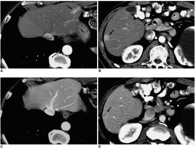
Fig. 12.
Tumorous (hepatocellular carcinoma) and nontumorous arterioportal shunt in cirrhotic liver.
A, B. CT scans obtained during hepatic arterial phase depict focal hyperattenuating lesions in segment II (arrow in (A)) and segment VI (arrow in (B)). Both lesions are rectangular-shaped and located in subcapsular areas. Note the enhanced peripheral portal vein (arrowheads in (A)) inside the lesion in segment II.
C, D. Portal venous phase images show no lesion in segment II, indicating that the lesion depicted in (A) is a nontumorous arterioportal shunt; however, a nodular hypoattenuating lesion with rim enhancement is seen in segment VI (arrow in (D)), indicating hepatocellular carcinoma. Note that the size and shape of the lesion in segment VI differ slightly between the hepatic arterial phase (B) and portal venous phase (D) images, indicating the presence of arterioportal shunt around the tumor.

