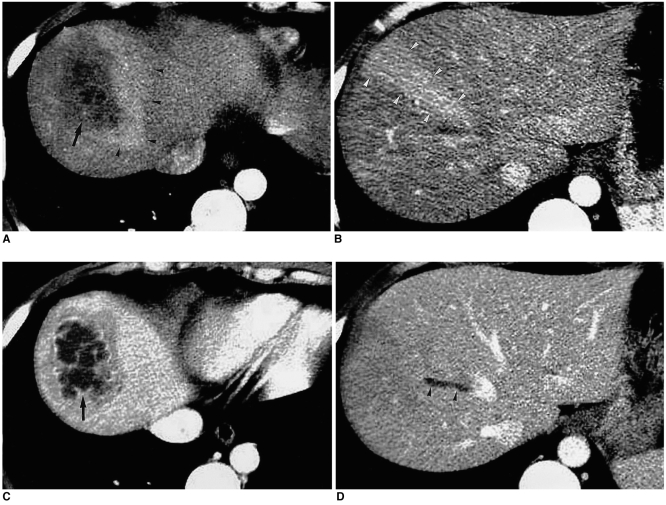
Fig. 13.
Arterioportal shunt secondary to hepatic abscess.
A, B. CT scans obtained during hepatic arterial phase show a hepatic abscess (arrow), with wedge-shaped hyperattenuation (arrowheads) in the adjacent parenchyma.
C, D. Portal venous phase images depict abscess wall enhancement, and the demarcation of the abscess is clearer than in the hepatic arterial phase image (arrow). Portal venous phase images show that parenchymal enhancement has returned to normal. Note that the wedge-shaped parenchymal hyperattenuation demonstrated by hepatic arterial phase imaging points to the thrombosed hepatic vein (arrowheads).

