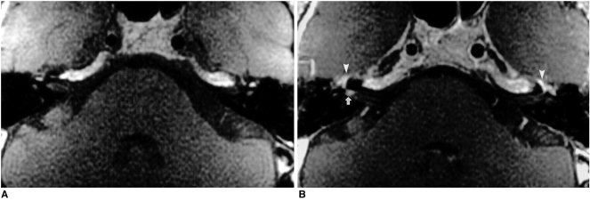
Fig. 1.
A 42-year-old man with right-sided Bell's palsy.
Axial pre- (A) and postcontrast (B) T1-weighted MR images demonstrate focal enhancement of the right facial nerve at the fundus of the internal auditory canal (arrow). Note the symmetric, intense enhancement of the facial nerves around the geniculate fossa on both sides (arrowheads), attributable to the prominent normal circumneural arteriovenous plexus located in this area.

