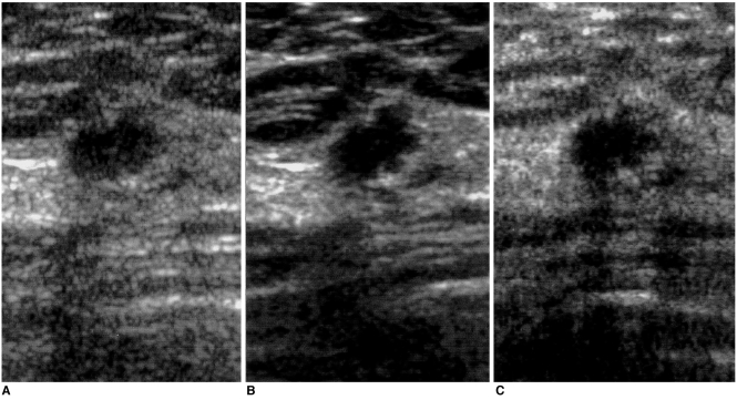
Fig. 1.
Sonograms of a 56-year-old woman show an oval-shaped, microlobulated and spiculated marginated, hypoechoic nodule. In lesion conspicuity and margin depiction, real-time compound (B) and pulse-inversion harmonic imaging (C) are better than conventional scanning (A). Pathologic examination showed that the case was invasive ductal carcinoma.

