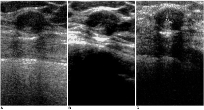
Fig. 4.
Sonograms of 32-year-old woman reveal a well-defined, oval-shaped, heterogeneous isoechoic nodule. The heterogeneous internal echotexture is more clearly visualized by real-time compound imaging (B) than conventional (A) and pulse-inversion harmonic imaging (B) and an internal microcalcification (curved arrow) is revealed by pulse-inversion harmonic imaging (C). Pathologic examination showed that the condition was atypical ductal hyperplasia.

