Fig. 5.
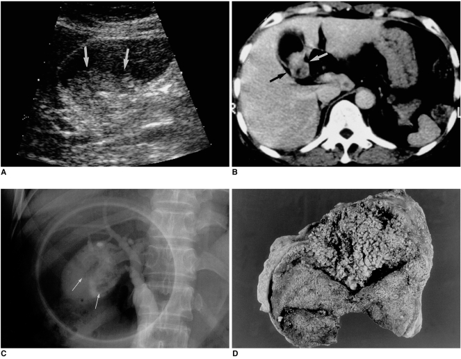
A 56-year-old woman with papillary adenomatosis in which multifocal foci of carcinomatous change are seen in the gallbladder and cystic duct.
A. Sonogram of the gallbladder reveals an echogenic mass with a ragged surface (arrows).
B. Post-contrast CT image depicts an enhancing mass (arrows) in the gallbladder.
C. Endoscopic retrograde cholangiogram indicates that the gallbladder mass has a nodular and velvety appearance (arrows).
D. Photograph of resected gallbladder depicts an irregular mass, velvety in appearance, comprised of myriads of papillary projections.

