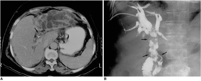
Fig. 7.
A 59-year-old woman with papillary carcinomatosis in the left hepatic ducts and extrahepatic ducts.
A. CT image of the liver during the equilibrium phase shows markedly dilated, tortuous, crowded bile ducts in the left hepatic lobe. The percutaneous transhepatic catheter present in the extrahepatic duct is displaced posteriorly due to the intraluminal mass (arrow).
B. Percutaneous transhepatic cholangiogram demonstrates multiple nodular filling defects in the extrahepatic ducts (arrows), simulating multiple stones. Due to obstruction, the left hepatic duct is not opacified.

