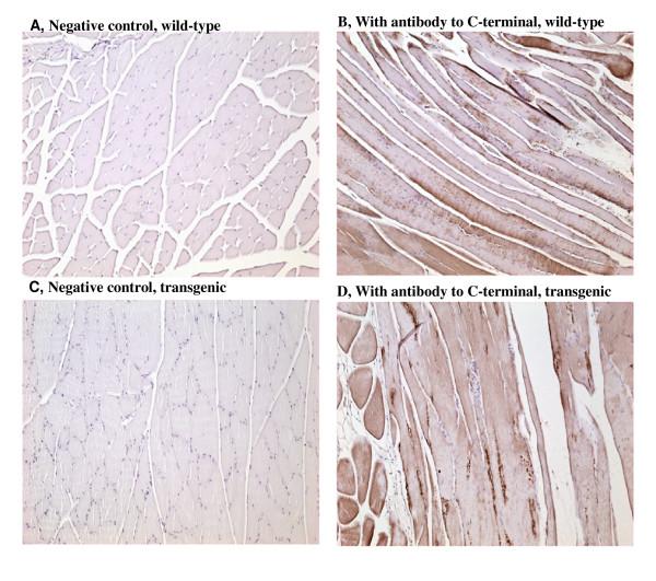
Figure 2.
Localization of myostatin in biceps femoris by immunohistochemistry with antibody to the C-terminal. Mice were sacrificed at 12 months of age. Biceps femoral muscle samples were used for this study. The cross-sectional area of the muscle samples was frozen-sectioned. The sections were blocked in 10% rabbit serum and incubated with or without (negative control, A and C) the myostatin C-terminal antibody, followed by incubation with an HRP-coupled secondary antibody and counterstaining with diaminobenzidine (DAB)/peroxidase reaction (B and D). Panel A and C show the myofibers which were cross-sectioned, and B and D show myofibers which were longitudinally sectioned. Nuclei were stained as purple/blue color, and myostatin protein was stained as brown color.

