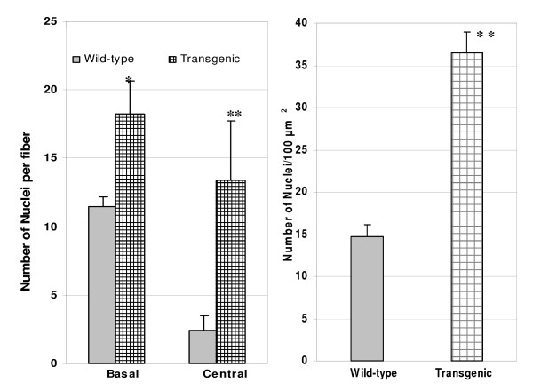
Figure 4.
Comparison of number of myofiber nuclei between wild-type and transgenic mice. Biceps femoral muscle samples of three transgenic and three wild-type littermate mice were used for this study. Ten random microscopic fields under 40× magnification were selected for both wild-type and transgenic mice. The number of nuclei in the center and the basal lamina for each muscle fiber bicep muscles were counted. The average number of nuclei per 100 um2 were calculated by an image analysis software. Bars represent mean ± SEM (n = 10 per group). Superscript ** and *above the bars indicate significant differences at P < 0.01 and P < 0.05, respectively.

