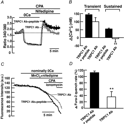
Figure 4. TRPC1 mediates CCE in mouse PASMCs.
A, TRPC1 antibody (1 : 100) inhibited the CPA-induced sustained but not transient increase in fura-2 fluorescence ratio in the presence of 10 μm nifedipine. B, bar graph showing mean changes in transient and sustained increase in [Ca2+]i caused by 10 μm CPA after re-addition of 2 mm Ca2+ in the presence of 10 μm nifedipine, in control cells (filled bars, TRPC1 Ab+peptide, n= 156) and in cells treated with TRPC1 antibody (open bars, n= 139). **P < 0.01 (unpaired t test). C, TRPC1 antibody (1 : 100) inhibited the increase in Mn2+ quench of fura-2 fluorescence caused by 10 μm CPA in the presence of 10 μm nifedipine. D, bar graph showing percentage change in fura-2 quench rate after store depletion in the presence of 10 μm nifedipine, in control cells (filled bar, TRPC1 Ab+peptide, n= 117) and in cells treated with TRPC1 antibody (open bar, n= 48). **P < 0.01 (unpaired t test).

