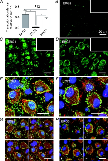
Figure 7. Immunohistochemistry shows both ERG1 and ERG3 are present in MNTB from P12 mice.
A, QRT-PCR of ERG1, 2 and 3 mRNA expressed relative to Kv3.1b from P12 mice (N= 3, n= 3). ERG1 and ERG3 were present at significantly higher levels than ERG2 (one-way ANOVA with Bonferroni's correction, *P < 0.05). B–D, fluorescence images showing ERG1, 2 and 3 antibody staining. B, ERG2 shows low staining levels. ERG1 (C) and ERG3 (D) immunoreactivity was high in the MNTB principal neurons. Insets show corresponding blocking peptide controls. E and F, confocal projections for ERG1 (green) (E) and ERG3 (green) co-labelled with Kv3.1b (red) and DAPI (blue) (F). G and H, four single optical z sections taken at 1 μm intervals through the same cells shown in E and F. Note the clear punctate membrane staining for both isoforms (arrowheads, Ga and Ha) and axon staining (arrowheads Gb and Hc). Scale bar (20 μm) applies to all panels.

