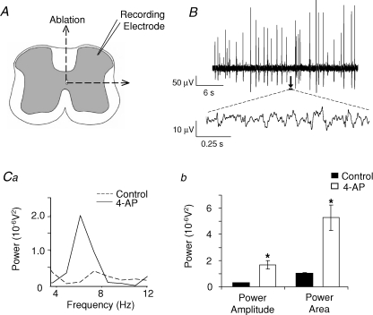
Figure 2. 4-AP evokes two profiles of activity in SG of isolated DH quadrants in vitro.
A, to isolate the DH from contralateral DH and ipsi- or contralateral ventral regions, intact spinal cord slices were lesioned vertically through the midline and horizontally just below lamina V/VI. B, 4-AP (50 μm) induced two profiles of activity similar to that observed in intact slices, namely large amplitude field population spiking activity (B, upper trace) and low amplitude rhythmic oscillations within the inter-spike interval (B, lower trace, sample expanded 1 s epochs indicated by arrow and black bar). Ca, power spectra of the low amplitude rhythmic oscillations reveal a peak 4–12 Hz frequency band with increased power amplitude and area (Cb) during 4-AP application (dashed line, control; continuous line, 4-AP).

