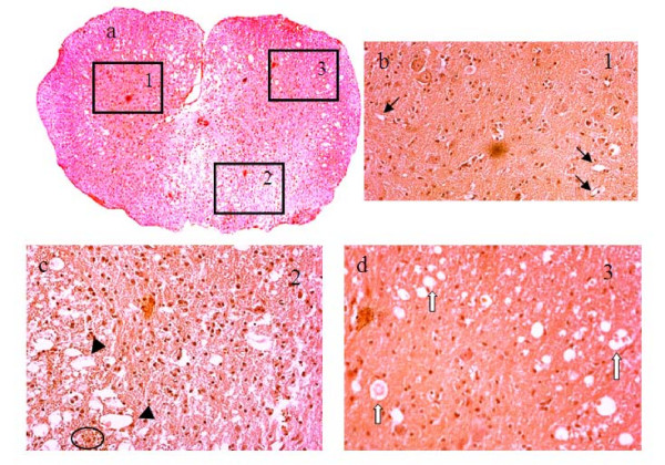
Figure 7.
H&E stained histology images of injured SC on day 3: a) at lower magnification and b-d) at higher magnification within the selected square windows marked with numbers 1, 2 and 3 in panel a. The windows were selected based on the MRI-observed pathology in Figure 6. The 1st window was selected in an intact GM region, the 2nd window was selected in a significantly damaged region and the 3rd window was selected in a region with edema. Black arrows denote normal vessels and capillaries. Black arrow heads point to disrupted vasculature with damaged BSCB. White arrows point to vessels surrounded by cavities. Circle denotes clusters of extravasated red blood cells.

