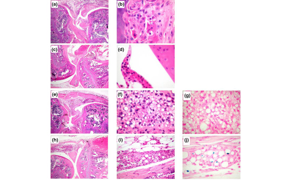
Figure 8.
Histology of mouse knee joints 4 days after intra-articular injection. Staining is with haematoxylin and eosin unless specified otherwise. (a, b) Antigen-induced arthritis (AIA), positive control. (a) Intense inflammatory infiltrate in the synovial tissue and the joint cavity. (b) At a higher magnification, mononuclear inflammatory cells destroyed cartilage and modulated bone. (c, d) Negative control, phosphate-buffered saline. No inflammatory infiltrate is present either in the synovial tissue or the joint cavity. The cartilage surface is smooth. (e-g) AIA knees treated with microparticles without iron or dexamethasone 21-acetate (DXM). (e) Pronounced inflammatory infiltrate and cartilage destruction by a synovial 'pannus'. (f) Presence of numerous microparticles in synovial macrophages mixed with some polynuclear cells. (g) Prussian blue (PB) staining without evidence of iron. (h-j) AIA knees treated with microparticles containing iron and DXM. A reduction of inflammation in the synovial tissue is apparent when compared with (e-g). (h) No inflammation of the joint cavity or cartilage invasion or bone destruction is apparent. (i, j) Presence of microparticles in macrophages of the synovial tissues containing iron (j, PB). Original magnifications: × 20 (a, c, e, h) and × 400 (b, d, f, g, i, j).

