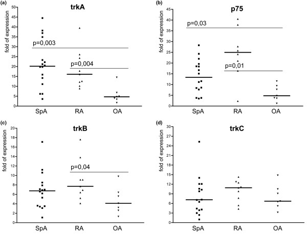
Figure 2.
Scatter plots showing mRNA expression of neurotrophin receptors. The scatter plots a to d depict the expression levels of the four neurotrophin receptors. (a) Tyrosine kinase (trk)A. (b) p75. (c) trkB. (d) trkC. Bold horizontal lines represent the median. The highest levels were found for trkA and p75, revealing significantly higher expression levels in spondyloarthritis (SpA; P = 0.0003, P = 0.003 respectively) and rheumatoid arthritis (RA; P = 0.004 and P = 0.001, respectively) vs osteoarthritis (OA).

