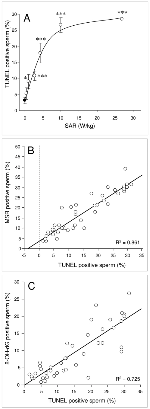
Figure 5. RF-EMR induces DNA fragmentation in human spermatozoa.
Following Percoll fractionation, 5×10⁶ high density spermatozoa were resuspended in 1 ml BWW, pipetted into 35 mm Petri dishes and placed inside a waveguide. 5×10⁶ cells in 1 ml BWW were placed outside the waveguide as a control (closed circle). The cells in the waveguide were exposed to 1.8 GHz RF-EMR at SAR levels between 0.4 and 27.5 W/kg (open circles) and all samples were incubated for 16 h at 21°C. Following incubation, cells were fixed; DNase-I was used as a positive control. After 1 h incubation at 37°C, 50 µl of label and enzyme master mixes were added to the cells and incubated for 1 h at 37°C. Cells were then washed and assessed by flow cytometry. A, Significant levels of DNA fragmentation was observed in exposed spermatozoa at 2.8 W/kg (*p<0.05) and above (***p<0.001). B, DNA fragmentation was positively correlated with ROS production by the mitochondria as monitored by MSR (R2 = 0.861). C, 8-OH-dG was also positively correlated with DNA fragmentation (R2 = 0.725). Results are based on 4 independent samples.

