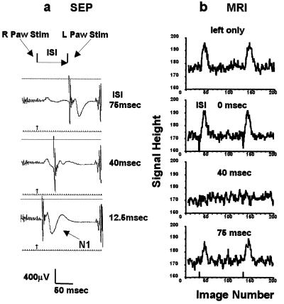
Figure 5.
Bilateral forepaw stimulation with time delay (Paradigm III). (a) SEP responses at the contralateral somatosensory area to the left forepaw. (Top) ISI 75 msec. (Middle) 40 msec. (Bottom) 12.5 msec. The noises at far right were EPI generated. The sharp spikes at the second stimulation were electrical artifacts. (b) The time courses of BOLD responses (without EEG electrodes) with varied ISI. The pair of stimulations was repeated four times at every 620 msec. Left paw stimulation only, from the top: ISI 0 msec, ISI 40 msec, and ISI 75 msec. The sharp spikes at the onset of stimulation with ISI = 0 msec were due to a small and transient head motion.

