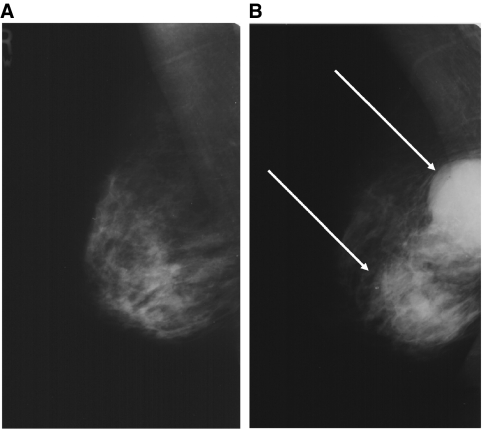
Figure 2.
(A) The mammogram of a 58-year old patient referred with an 8-week history of a clinically benign (E2) lump inferior to the right nipple, reported as normal at initial assessment and subsequent review. A breast ultrasound (US) of the palpable lump by a consultant radiologist found no suspicious abnormality and the patient was discharged. (B) Mammogram taken 2 years later when the patient re-presented stating that the lump had enlarged and clinical examination showed a clinically malignant (E5) abnormality, shows an obvious large bilobed mass (arrows) suspicious for malignancy, which was confirmed on US. A core biopsy showed a metaplastic carcinoma.

