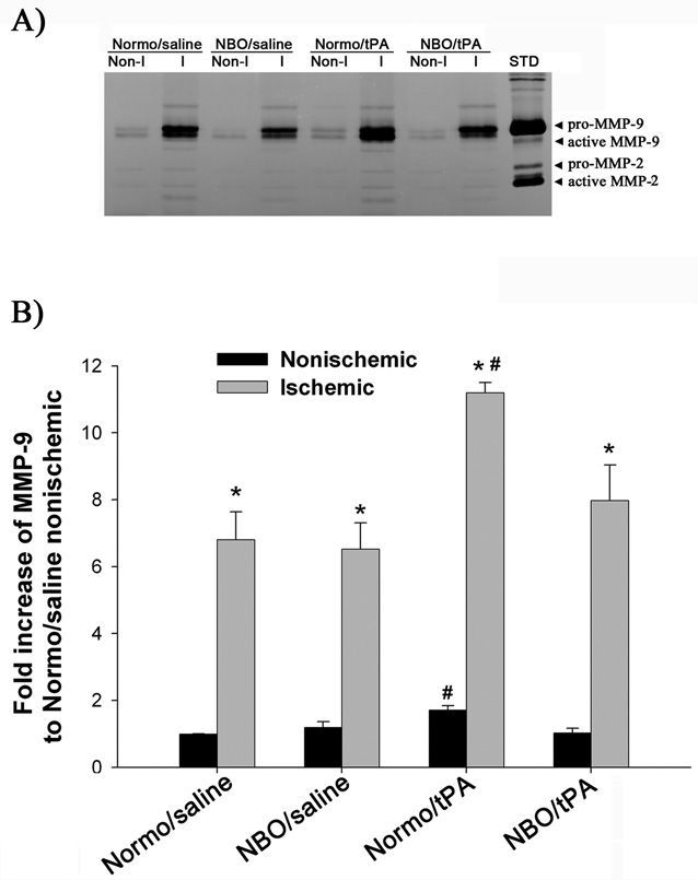
Figure 4.
Effects of NBO and tPA on MMP-9 induction in ischemic cerebral microvessels. A) Representative gelatin zymogram showing MMP-9 expression in the nonischemic (Non-I) and ischemic (I) hemispheric cerebral microvessels for each group. STD is a mixture of standard MMP-2 and MMP-9. B) Quantified MMP-9 band intensity for each group. A significant increase in MMP-9 levels was observed in the ischemic hemispheric microvessels in all animal groups (*p<0.05, versus Nonischemic; paired t-test). Normo/tPA rats showed higher levels of MMP-9 in both ischemic and nonischemic hemispheric microvessels than the other three groups (#P<0.05). Data are expressed as mean±SEM.

