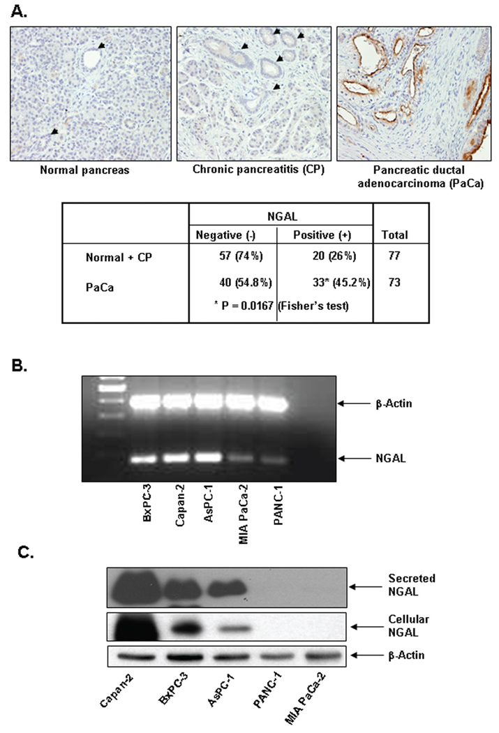
Figure 1. NGAL expression in PaCa tissue and PaCa cells.
A. Tissue microarrays from human PaCa (MDACC) were immunohistochemically stained by using a monoclonal NGAL antibody as described in Materials and Methods. The table shows a summary of NGAL IHC of the PaCa tissue microarrays. B. RT-PCR for NGAL expression by using NGAL-specific primers after total RNA extraction from PaCa cells as described in Materials and Methods. C. NGAL expression were determined by western blots of protein lysates or culture media from PaCa cells as described in Materials and Methods.

