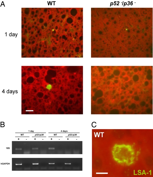
Fig. 3.
P. falciparum p52−/p36− parasites do not grow and persist in a humanized chimeric liver mouse model. SCID Alb-uPA mice were inoculated with 106 primary human hepatocytes to create chimeric human–mouse livers. Mice positive for engraftment were infected intravenously with 1 × 10⁶ sporozoites of either p52−/p36− or WT P. falciparum. (A) Immunofluorescent micrographs of chimeric SCID/Alb-uPA liver cryosections after WT P. falciparum infection, stained with parasite-specific antibodies. Tissue was harvested at 1 day or 4 days after mice were inoculated with P. falciparum sporozoites. Sections in panels were stained with an anti-PfHSP70 monoclonal antibody. The data show that p52−/p36− liver stages are detected 1 day after infection but are not detected 4 days after infection. (Scale bar: 10 μm.) (B) RT-PCR analysis was performed with RNA isolated from individual livers of chimeric mice 1 day or 4 days after infection. Total RNA was extracted, and reverse-transcription reactions were done with (+) or without (−) the reverse transcriptase. Primers specific for the 18S (small-subunit) ribosomal RNA of P. falciparum and human glyceraldehyde phosphate dehydrogenase (hGAPDH) were then used to amplify the reverse-transcribed message. The GAPDH control reaction was positive for samples from all of the mice. The parasite-specific 18S reaction was positive for the mice infected with the WT parasites at day 1 and day 4 after infection. For the double-knockout parasite-infected mice, the 18S reaction was only positive for tissues harvested 1 day after infection and negative for tissue harvested at 4 days after infection, indicating that p52−/p36− parasites were no longer present in the liver at the later time point. RT-PCR was performed on all mice used in the experiment, and results were consistent with the results shown here. (C) An example of WT P. falciparum liver stage from chimeric SCID/Alb-uPA liver cryosection stained with polyclonal rabbit antisera against the repeat region of LSA-1 at 4 days after infection. (Scale bar: 5 μm.)

