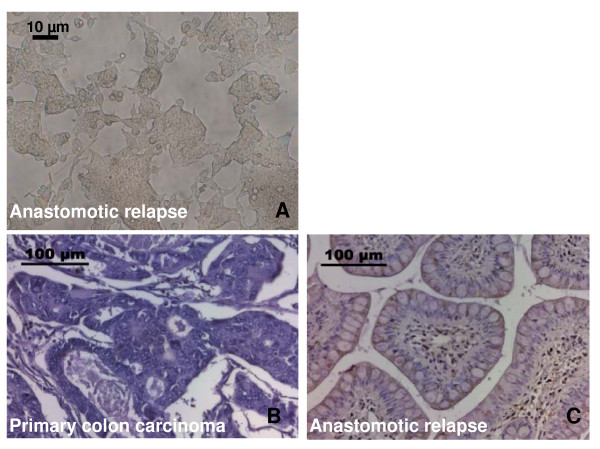
Figure 2.
A- Photomicrograph view of the patient-derived cell line of the anastomotic relapse. Cells were cultured and passaged twice a week. The picture was taken at sub-confluent stage at cell passage 26; the scale bar marks 10 μm. B/C: Comparative immunohistochemical analysis of the cytosolic Hsp70 content in the primary colon carcinoma (B) and the anastomotic relapse (C). Histological slides were stained with the Hsp70 specific antibody 3B3 which reacts with Hsp70 and does not cross-react with Hsc70; the scale bar marks 100 μm.

