
Figure 2.
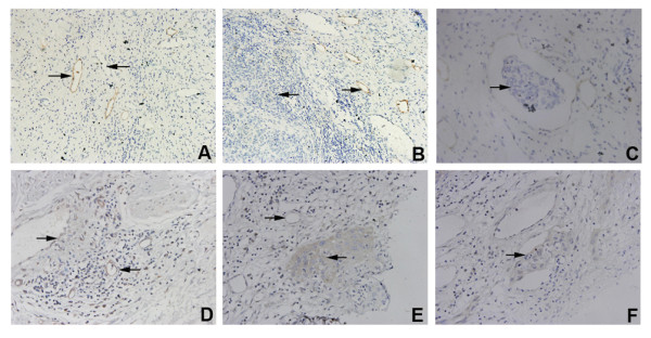
Morphological features of LYVE-1 positive lymphatic vessels and Flt-4 positive vessels in cervical cancer tissues. A. The LYVE-1 positive lymphatic vessels (→) were clearly different from blood vessels (←) ×200; B. The LYVE-1 positive lymphatic vessels (→) were mainly distributed in the paratumor stromal tissue (←) ×400; C. Some LYVE-1 positive lymphatic vessels contained invaded tumor cells (→) ×400; D. Some Flt-4 positive vessels were similar to blood vessels in their morphology (→), and others were similar to lymphatic vessels (←) ×400; E. The Flt-4 positive vessels (→) were mainly distributed in the paratumor stromal tissue (←) ×400; and F. Some Flt-4 positive vessels contained invaded tumor cells (→) ×400.