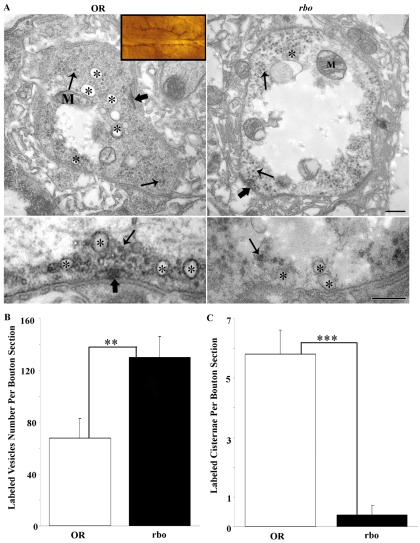
Fig. 10.
RBO is required for activity-dependent endosome formation in synapses. FM1-43 photoconversion generates an electron-dense marker that is clearly visible by electron microscopy. Dye was incorporated into NMJ synapses after a 2-minute high-[K+] stimulation at 37°C, photoconverted and examined in wild type (OR) and rbots. (A) Inset shows a light-microscopy image of muscle 6/7 NMJ as photoconverted, ready to be sectioned at the electron-microscopic level. Top panels show representative images of control and mutant bouton profiles. Many FM-labeled vesicles and endosomes can be seen throughout the boutons. Asterisks (*) mark labeled endosomes in wild type; note the absence of label in rbots. Lower panels show higher-magnification images of endosomes (*) and SVs (thin arrows). In rbots, no labeling of endosomes was observed (*), but abundant SVs were labeled. Scale bars: 250 nm. (B) Quantification of FM-labeled SVs per bouton section. (C) Quantification of FM-labeled endosomes per bouton profile. Significance indicated: **P<0.01, ***P<0.001.

