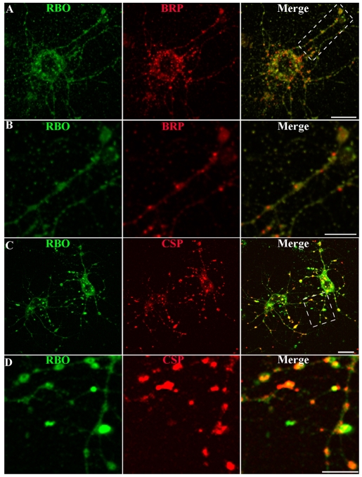
Fig. 3.
RBO localizes to synapses in central brain primary neuron cultures. Representative images of neuron cultures of rbo2/rbo2; rbo-egfp/rbo-egfp. (A) RBO-GFP (green) at 6 DIV, double-labeled with anti-BRP (red) marking active zones. Scale bar: 10 μm. (B) Area in boxed area in A magnified to show RBO-GFP and BRP colocalization in axonal synaptic varicosities. Scale bar: 5 μm. (C) RBO-GFP (green) double-labeled with anti-CSP (red) marking synaptic punctae. Scale bar: 10 μm. (D) Area in boxed area in C magnified to show RBO-GFP and CSP colocalization in synaptic punctae. Scale bar: 5 μm.

