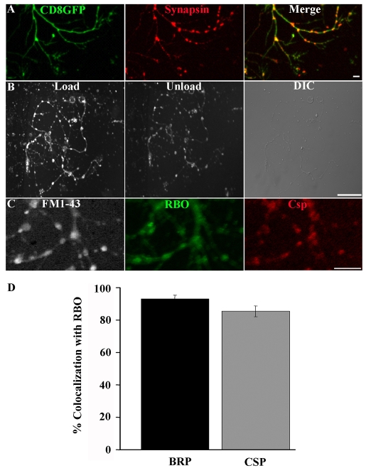
Fig. 4.
RBO localizes to functional synapses with cycling SVs. (A) Cultured neurons containing elav-GAL4 driven UAS-CD8::GFP (green) doubled-labeled with anti-Synapsin (red) to show axonal varicosities containing the presynaptic marker. Scale bar: 2 μm. (B) FM1-43 labeling with 60 mM [K+] for 45 seconds loads synaptic varicosities (left). A shorter, 30 second, exposure to 60 mM [K+] partially unloads dye (middle). Nomarski DIC image showing neuronal structure (right). Scale bar: 20 μm. (C) In rbo2/rbo2; rbo-egfp/rbo-egfp 6-DIV cultures, loaded FM1-43 dye (white) colocalizes with RBO-GFP (green) and the synaptic marker anti-CSP (red). Scale bar: 5 μm. (D) Quantification of the percentage of colocalization of BRP and CSP with RBO-GFP punctae.

