Fig. 7.
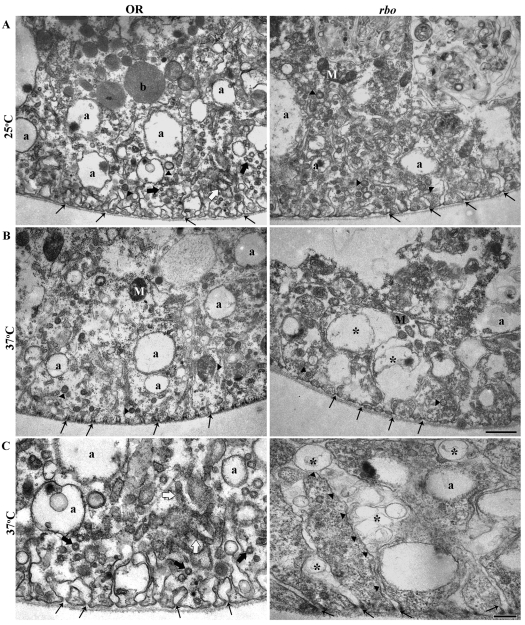
Ultrastructure of Garland cells in wild type and in rbots mutants. (A) Transmission electron microscopy (TEM) images of wild-type and rbots Garland cells at 25°C. The cortical cell region extends ∼3 μm, with many labyrinthine channels (thin black arrows). Numerous α vacuoles (a), longitudinal (white arrows) and transverse (thick black arrows) tubular elements, and coated vesicles and/or pits (arrowheads) are present in all section planes. Many sections also contain mitochondria (M) and β vacuoles (b). Wild type and rbots appear indistinguishable at 25°C. (B) Following 10 minutes at 37°C, the number of α vacuoles in rbots cells is reduced, with remnant vacuoles being larger and more irregular. Labyrinthine channels swell and distend with many vacuolar structures (*). Scale bar: 1 μm. (C) Higher-magnification images at 37°C. In rbots, very elongated labyrinthine channels extend into the cell; these channels contain many vacuolar structures (*). Thin black arrows show the origin of the labyrinthine channels at the basement membrane. Scale bar: 250 nm.

