Fig. 8.
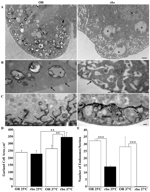
Block in HRP endocytosis occurs in Garland cells in the absence of RBO. (A) Garland-cell endocytotic activity visualized using HRP uptake in wild type (OR) and rbots after 10 minutes at 37°C. Many coated profiles of α vacuoles (a) are labeled in wild type (left), among pre-existing unlabeled α vacuoles (arrows). The mutant (right) shows no HRP uptake into α vacuoles (a). N, nucleus. Scale bar: 1 μm. (B) Higher-magnification images of α vacuoles (a) and labyrinthine channels (arrows). The mutant (right) shows an absence of labeling, with distended and swollen labyrinthine channels. Scale bar: 250 nm. (C) Tannic-acid impregnation shows labyrinthine channels that are continuous with the extracellular space. In rbots, the channels are fused with many vacuolar inclusions. Scale bar: 250 nm. (D) Quantification of cell area. No change was observed in rbots at 25°C compared with wild type, but a highly significant increase in cell area was seen at 37°C. (E) Quantification of loaded endosomes per section. A significant reduction in the number of loaded endosomes was observed in rbots at 25°C compared with wild type, with near complete loss at 37°C.

