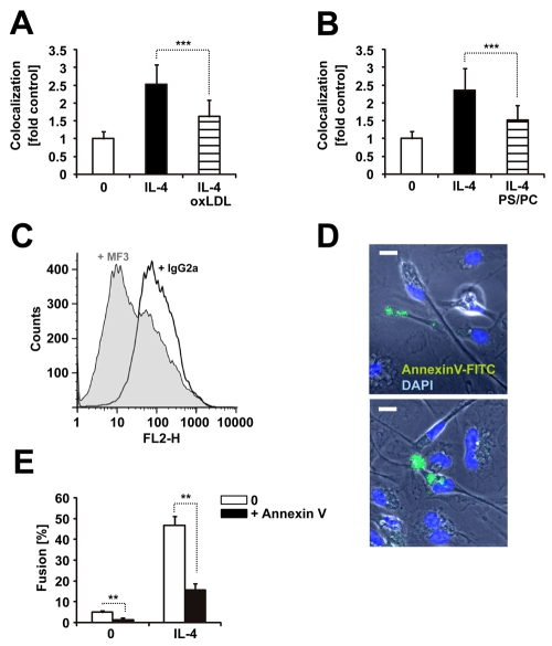
Fig. 6.
A role for lipid recognition by CD36 and PS exposure and recognition during macrophage fusion. Quantitation of ThioMΦ fusion in the presence of (A) oxLDL (50 μg/ml) or (B) PS liposomes (PS/PC, 50 μM). Means±s.d. combined from three independent experiments. (A) n=21, ***P<0.0001; (B) n=17, ***P=0.0003. Mann Whitney Test, two-tailed. (C) Binding of DiI-labelled PS liposomes (=FL2) to ThioMΦ can be blocked by addition of anti-CD36 antibodies (MF3) but not control antibodies (IgG2a). This result was obtained in three independent experiments. (D) ThioMΦ were plated with IL-4 on Permanox in the presence of annexinV-FITC for 18-24 hours. Nuclei were counterstained with DAPI. (E) Annexin V-FITC blocks IL-4 induced ThioMΦ fusion. Means ±s.d., n=6, **P=0.0051, Mann Whitney Test, two-tailed. Shown are representatives of three independent experiments. Bar, 10 μm.

