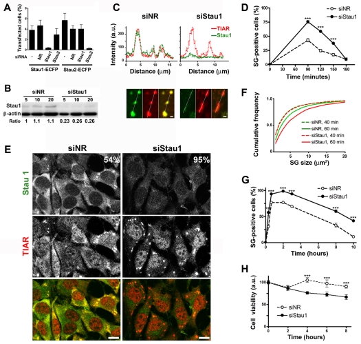
Fig. 3.
Stau1 depletion facilitates stress granule formation. (A) Stau1-ECFP or Stau2-ECFP constructs were independently transfected into COS-7 cells simultaneously with the indicated siRNAs (NR, non-relevant). Expressing cells and total cells identified by DAPI staining were counted 16 hours after transfection. (B) NIH 3T3 cells were treated with siNR or siStau1 and extracts were analyzed by western blot. Intensity of the Stau1 signal relative to that of β-actin indicates a 75% reduction of Stau1 levels. (C) Line-scan analysis of arsenite-induced stress granules in siNR- or siStau1-treated cells indicates the presence of negligible amounts of Stau1 upon Stau1 depletion. Scale bar: 1 μm. (D) Upon treatment with the indicated siRNAs, cells were continuously exposed to arsenite and cells containing stress granules (SGs) were identified by TIAR staining. A representative experiment out of three is shown where approximately 200 siNR-treated cells and 300 siStau1-treated cells randomly selected from duplicate coverslips were analyzed for each time point. Stress granule formation is facilitated in Stau1-depleted cells; ***P<0.0001 for each data pair. (E) TIAR and Stau1 staining in siNR- and siStau1-treated cells after 1 hour arsenite treatment. Percentage of cells containing stress granules is indicated. (F,G) After treatment with the indicated siRNA, NIH 3T3 cells were exposed to thapsigargin and stained for TIAR and Stau1. (F) stress granule size was evaluated in approximately 1800 stress granules present in 90 cells at 40 or 60 minutes after thapsigargin treatment. (G) Time course of stress granule formation. A representative experiment out of three is depicted. A minimum of 400 cells was analyzed for each point. Stau1 depletion significantly facilitates the formation of stress granules induced by ER stress (***P<0.0003). (H) NIH 3T3 cells were treated with the indicated siRNAs and continuously exposed to 100 nM thapsigargin. Percentage of viable cells was determined in triplicate using the MTT viability assay. A representative experiment out of three is depicted. ***P<0.0001.

