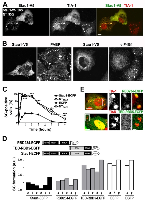
Fig. 4.
Stau1 overexpression inhibits stress granule formation. (A,B) COS-7 cells were transfected with Stau1-V5 and exposed to arsenite 24 hours after transfection. Representative cells showing the presence of stress granules in non-expressing or low expressing cells and their absence upon moderate Stau1-V5 overexpression are shown. Percentage of transfected or non-transfected cells containing stress granules is indicated. Scale bars: 10 μm. (C) NIH 3T3 cells were continuously exposed to thapsigargin 16 hours after transfection with Stau1-ECFP or ECFP. Cells with moderate level of expression (less than four times the endogenous levels) were analyzed for stress granule formation. A minimum of 120 transfected cells or neighboring non-transfected cells (NTECFP and NTStau1) from duplicate coverslips were analyzed. ***P<0.0001 for Stau1-ECFP expressing cells. (D) U2OS (a-c); NIH 3T3 (d); H1299 (e) Cos7 (f) or HeLa cells (g) were transfected with the indicated Stau1 construct and exposed to 250 nM or 500 nM thapsigargin (a and b); or to 0.1, 0.2 or 0.25 mM arsenite (c, d and e, respectively); or to 0.5 mM arsenite (f and g). Approximately 200 transfected and non-transfected cells were counted in each case. Stress granule formation is expressed as the ratio of the percentage of transfected stress granule-forming cells relative to that of neighboring non-transfected cells. The incidence of stress granules in non-transfected cells was as follows: a, 32%; b, 54%; c, 41%, d, 86%; e, 35%; f, 97% and g, 86%. Differences between averaged independent experiments (two in b and f) or between replicate coverslips (a-g) were less than 10% in all cases. (E) U2OS cells transfected with the indicated constructs were exposed to thapsigargin (upper panel) or arsenite (bottom panel). Representative stress granules showing inclusion of RBD234-EGFP or exclusion of TBD-RBD5-EGFP are shown. Scale bars: 10 μm (left panels) and 2 μm (insets).

