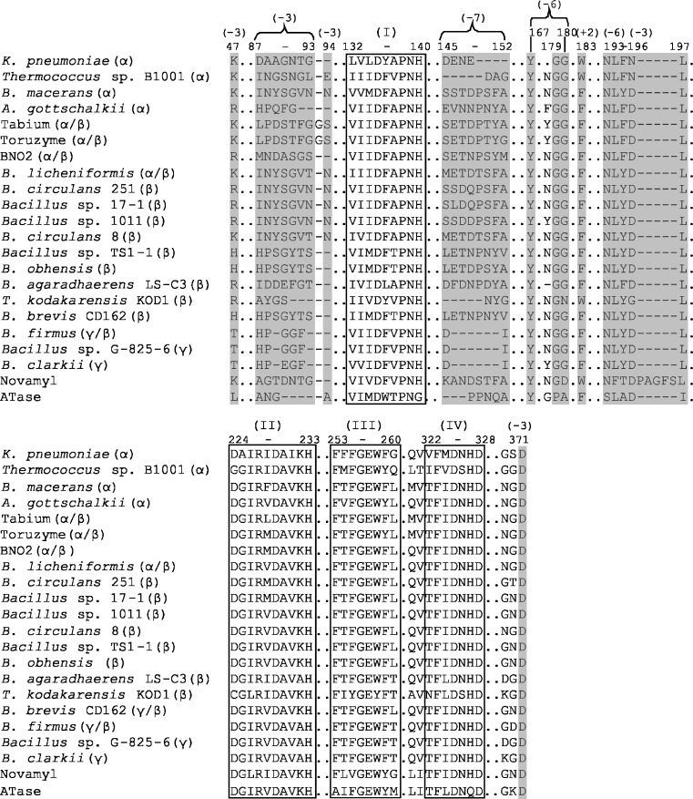
Fig. 3.
Alignment of functional CGTase regions. The four conserved GH13 sequence regions are boxed and marked I–IV. The numbers in brackets indicate the residues involved in substrate binding at subsites +2, −3, −6 and −7 and are coloured in grey. Numbering of residues according to BC251 CGTase. (See Fig. S1 of the Electronic supplementary material for full sequence alignment)

