Figure 1.
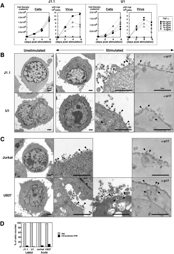
Reactivation of latently infected J1.1 and U1 cells displays HIV particle budding at the PM. (A) HIV production from J1.1 and U1 cells upon TNF-α stimulation. J1.1 and U1 cells were stimulated with TNF-α (~100 ng/ml). Levels of particle production were measured by p24 antigen ELISA. (B) HIV particle budding from J1.1 and U1 cells upon TNF-α stimulation. J1.1 and U1 cells stimulated with 50 ng/ml TNF-α were subjected to conventional electron microscopy and immunoelectric microscopy using anti-HIV-1 p17MA antibody. (C) HIV particle budding from acutely infected Jurkat and U937 cells. Jurkat and U937 cells were infected with HIV-1 (LAV strain) corresponding to 100–200 ng of p24CA antigen and were analyzed by electron microscopy. Arrowheads indicate budding particles and arrows indicate particles into intracellular vesicles in (B) and (C). (D) Semi-quantification of HIV-1 particle localization. Approximately 300 of particle-positive cells observed by conventional electron microscopy were sorted into the categories indicated.

