Figure 3.
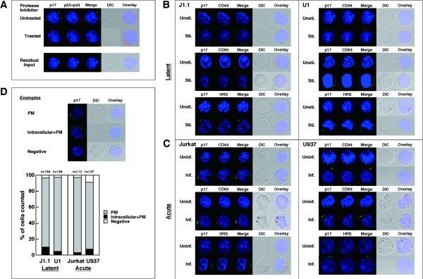
Upregulated molecules are accumulated to sites of HIV-1 particle budding. (A) Inhibition of Gag processing in J1.1 cells stimulated but treated with 1 μM ritonavir (upper) and residual HIV in Jurkat cells after infection (lower). For confocal microscopy, the cells were stained with anti-HIV-1 p17MA (green), p24CA (red) antibodies and with TOPRO-3 (blue). (B) Intracellular localization of upregulated molecules upon reactivation. J1.1 and U1 cells were either unstimulated (Unsti.) or stimulated (Sti.) with TNF-α and were immunostained with anti-p17MA antibody (green) and antibodies for CD44, CD63, and HRS (red). (C) Intracellular localization of upregulated molecules upon acute infection. Jurkat and U937 cells were infected with HIV-1 and immunostained. Inf., infected; Uninf., uninfected. (D) Semi-quantification of sites for HIV-1 particle production. Examples of cells exhibiting PM staining alone, intracellular+PM accumulations, and no signals (negative) (upper). Based on p17MA localization (PM, intracellular+PM, or negative), approximately 100–150 cells were sorted into the categories (lower).

