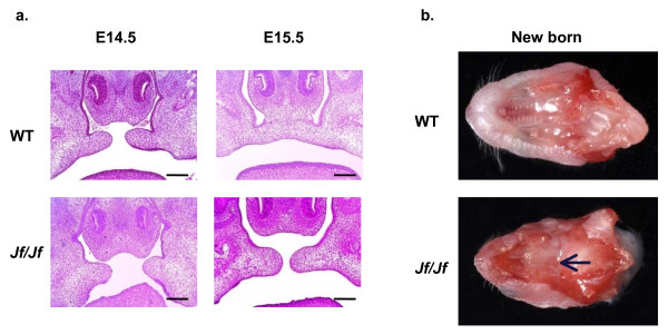
Figure 1.
Cleft palate phenotype. a. Coronal sections through the palate of E14.5 (before the fusion) and E15.5 (after the fusion) wild-type (WT) and homozygote (Jf/Jf) embryos, haematoxylin-eosin stained. Scale bars 200 μm. b. Cross-sections of heads showing secondary palate of a wild-type (WT) newborn mouse with fused palate and a homozygote (Jf/Jf) newborn mouse with a cleft (arrow).

