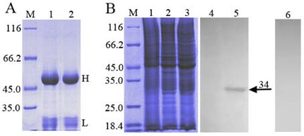
Figure 1.
(A) SDS-PAGE analysis of the purified UL51 antiserum and pre-immune serum. The purified IgG proteins of UL51 antiserum and pre-immune serum were respectively examined by SDS-PAGE (lanes 1 and 2). Molecular mass markers (in kDa) are shown to the left (lane M). H and L respectively indicate the position of heavy and light chains of the IgG proteins. (B) Reactivity and specificity of the purified UL51 antiserum analyzed by western blotting. DEF were mock-infected (lanes 1 and 4) or infected with DEV CHv strain (lanes 2, 3, 5 and 6) and harvested at 24 h p.i. The pUL51 was separated by SDS-PAGE (lanes 1 to 3) and analyzed by western blotting using the UL51 antiserum (lanes 4 and 5) or the pre-immune serum (lane 6). Molecular mass markers (in kDa) are shown to the left (lane M). The arrowhead indicates the position of the pUL51 (about 34 kDa).

