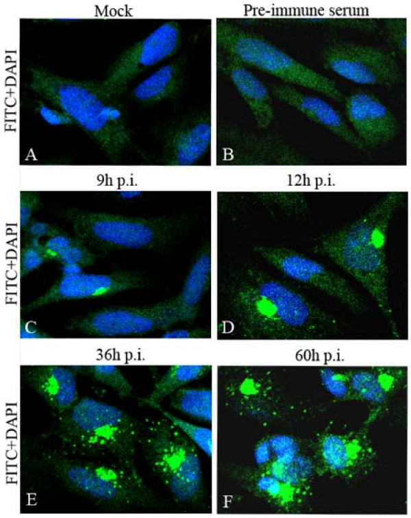
Figure 2.
Intracellular location and distribution of DEV pUL51 analyzed by IIF. Mock-infected (A) and DEV-infected (B to F) DEF were fixed as described in Materials and Methods. The samples were stained with the UL51 antiserum (A, C to F) or pre-immune serum (B), and reacted with anti-rabbit IgG-conjugated FITC, and then counter-stained with DAPI (blue is representative the cell nuclei). The merged fluorescence microscopy images of DEF are shown in panels A to F with high magnification (600×).

