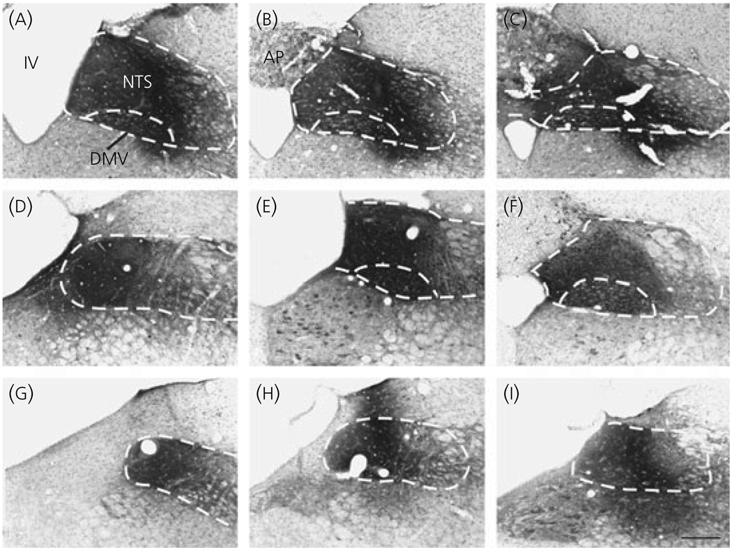
Fig. 3.
Photomicrographs of rostro-caudal sections of the dorsal vagal complex (illustrated by dotted lines), showing the location of the injection sites in three animals. The core of the cholera toxin B subunit injection sites covers the nucleus tractus solitarius (NTS) and the dorsal motor nucleus of vagus (dmv) in two animals (a–c and d–f), and confined to the boundaries of the NTS in a third animal (g–i). AP, Area postrema; IV, fourth ventricle; Scale bar = 200 µm.

