Fig. 7.
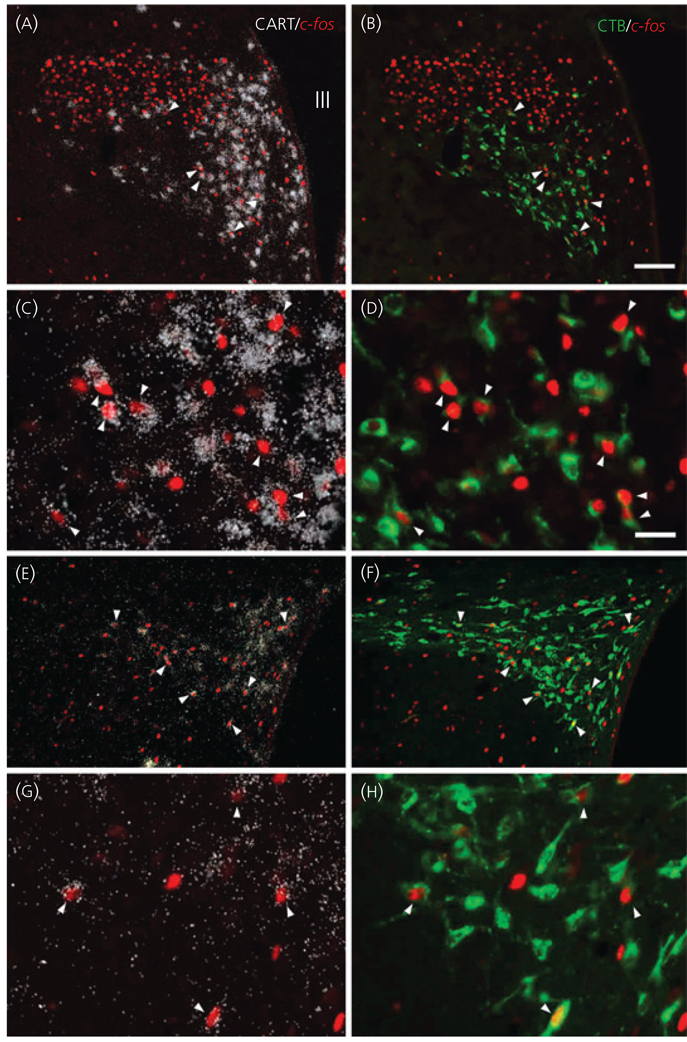
Location of cocaine- and amphetamine-regulated transcript (CART)-synthesising neurons in the hypothalamic paraventricular nucleus (PVN) that project to the dorsal vagal complex [CART/cholera toxin B subunit (CTB)/c-fos-immunoreactive (IR)] and express c-fos following lipopolysaccharide administration. Pairs of images of the same field under low (a,b) and high (c,d) magnification at the mid level of the PVN demonstrate triple-labelled neurons (arrowheads) containing CART mRNA (white silver grains), c-fos-IR (red immunofluorescence) nuclei, and retrogladely transported tracer CTB (green immunofluorescence) in the ventral parvocellular subdivision and in the ventral part of the medial parvocellular subdivision. Low (e,f) and high (g,h) magnification images of the caudal level of the PVN demonstrate c-fos containing CTB/CART neurons in the medial and lateral parvocellular subdivisions. Scale bars: 100 µm in (b) corresponds to (a), (e) and (f); 25 µm in (d) corresponds to (c), (g) and (h).

