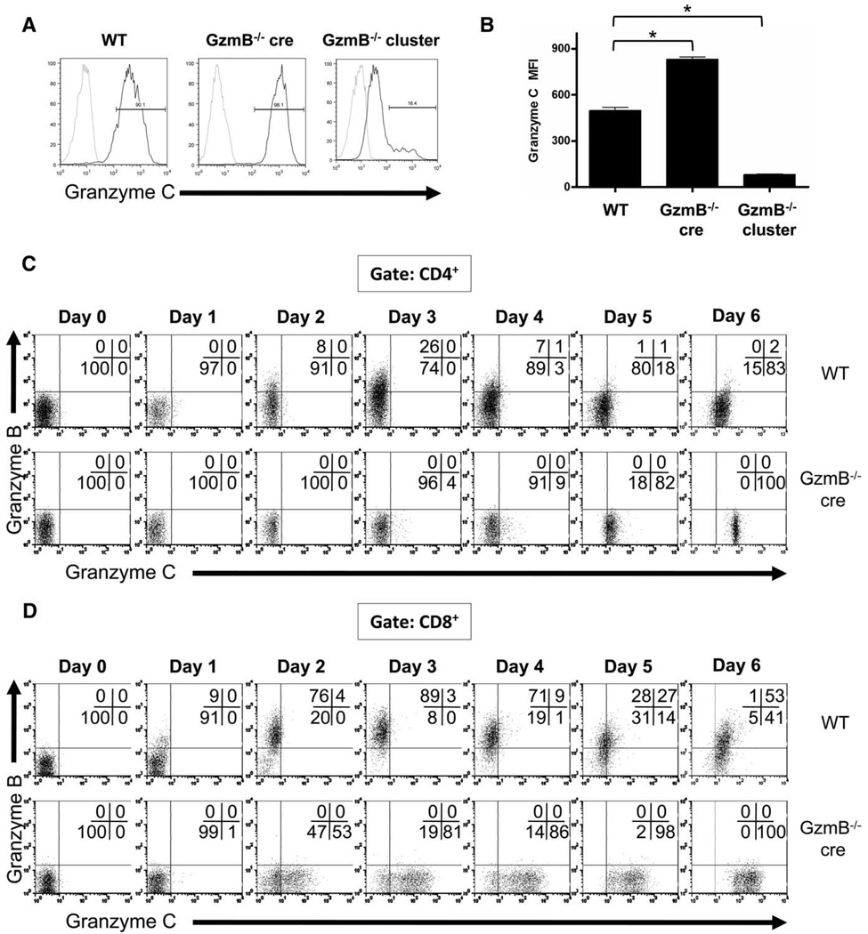
FIGURE 1.
Characterization of granzyme protein expression in activated lymphocytes using a novel granzyme C-specific mAb. LAK cells were generated by culturing splenocytes from WT, granzyme B−/− cre, or granzyme B−/− cluster-deficient mice in K10 medium supplemented with high-dose IL-2 (1000 U/ml). After 10 days of culture, LAK cells were harvested, fixed, permeabilized, and stained for intracellular granzyme C followed by staining with a PE-conjugated anti-hamster IgG secondary Ab. A secondary Ab alone condition (gray) was included as a negative control. Representative histograms are shown in A. A summary graph plotting MFI of granzyme C expression (mean ± SD) from three independent experiments is shown in B. *, p < 0.0001. WT and granzyme B−/− cre splenocytes were cultured in K10 medium with CD3/CD28 beads and harvested for flow cytometric analysis at various times during activation. T cell expression of granzymes B and C are shown. Results from an individual experiment are shown, gating on CD4+ T cells (C) and CD8+ T cells (D).

