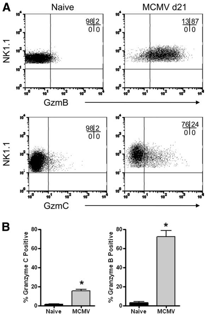
FIGURE 9.
NK cells express granzyme C protein in vivo following persistent MCMV infection. Rag1−/− C57BL/6 mice were infected with MCMV (2 × 10⁴ PFU i.p.) and splenic NK cells were evaluated for granzyme B and C protein expression at day 21 after infection. Representative flow plots from control littermates and day 21 postinfection Rag1−/− mice, gated on NK1.1+ cells, are shown in A. In the top panel, a PerCP-Cy5.5-NK1.1- conjugated Ab was used, and in the lower panel, an allophycocyanin-NK1.1 conjugate was used. The percentage of NK cells positive for granzyme B and C (mean ± SD) is summarized from three independent experiments in B. In total, six uninfected mice and nine MCMV-infected mice were analyzed. *, p < 0.05.

