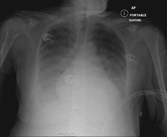
Figure 2.

Frontal chest radiograph showing features of alveolar pulmonary edema. The findings include opacification of both lungs with increasing density towards the lung bases due to a combination of air space shadowing and pleural effusions, cardiomegaly, upper lobe blood diversion (unreliable on supine AP radiograph) and an air bronchogram in the right upper zone
