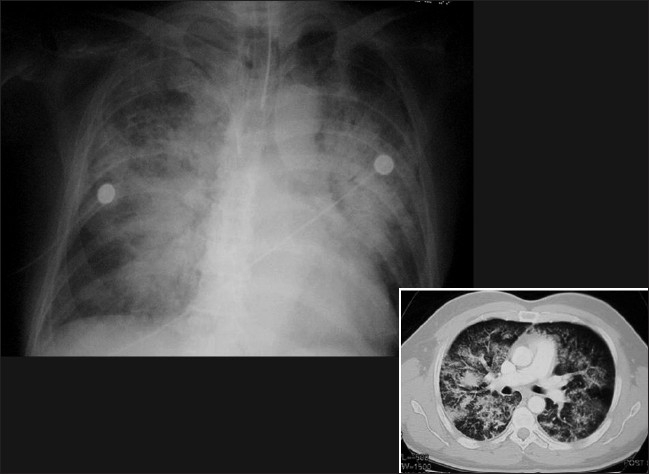
Figure 3.

A frontal chest radiograph and axial CT show features of ‘batwing’ alveolar pulmonary edema. Chest radiographic findings include bilateral opacities that extend in a fan shape outward from the hilum in a batwing; pattern. With worsening alveolar edema, the lung opacification becomes increasingly homogenous
