Figure 9.

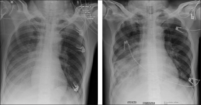
Same patient as in Figure 8; changes develop rapidly initially as mild opacification at the right lung base followed by lung parenchymal infiltrate associated with a small pleural effusion

Same patient as in Figure 8; changes develop rapidly initially as mild opacification at the right lung base followed by lung parenchymal infiltrate associated with a small pleural effusion