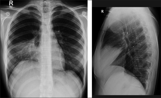
Figure 14.

Right–middle-lobe atelectasis may cause minimal changes on an AP supine chest radiograph. Note the loss of definition of the right heart border. A collapsed right middle lobe is more clearly defined on lateral radiograph, which is not commonly available in the ICU patient. Attention to the fissures reveals that the horizontal and lower portions of the major fissures move towards each other resulting in a wedge of opacity pointing to the hilum. This is a middle-lobe consolidation mimicking middle-lobe atelectasis
