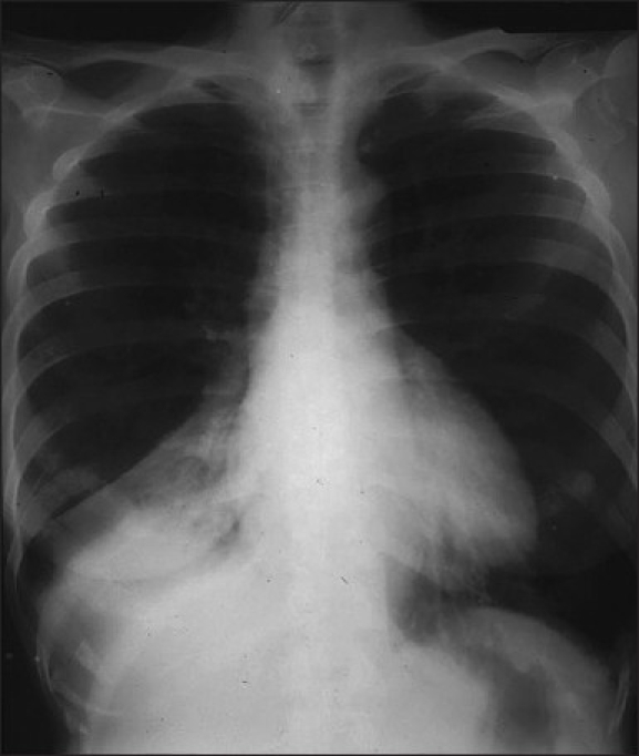
Figure 15.

An AP chest radiograph showing atelectasis of the right lower lobe. Note that the collapsing lobe has moved centrally and inferiorly towards the lower dorsal spine, where it is seen as a triangular opacity partially silhouetting the right hemidiaphragm and associated with a subtle air bronchogram. The minor fissure shows inferior displacement. Right–lower-lobe atelectasis can be differentiated from right–middle-lobe atelectasis by the persistence of the right heart border as in this case
