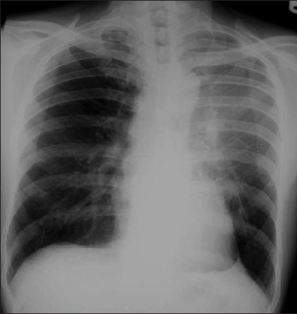
Figure 17.

A frontal chest radiograph showing a left–upper-lobe atelectasis. The radiograph reveals hazy opacification of the left hilum, elevation of the left hilum, near-horizontal course of the left main bronchus, posterior leftward rotation of the heart and the Luftsichel or air crescent sign, the name given to the appearance of aerated lung abutting the arch of the aorta, between the mediastinum and the collapsed left upper lobe. An appearance on a lateral radiograph, if available, of the ICU patient may show retrosternal opacity and displacement of the greater fissure anteriorly
