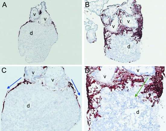
Fig. 1.
Double tissue confrontation assay using placental tissues at a gestational age of 6 weeks. Cryosections were stained with the monoclonal antibody MEM-G9 to visualize HLA-G, a specific marker for extravillous trophoblast. (A,B) Placental villous explants (v) firmly attach to the re-epithelialized decidual tissue (d). Outgrowing extravillous trophoblasts do not invade decidual tissues but rather only migrate on top of the decidual epithelium (blue arrows in B). (C,D) Placental villous explants (v) firmly attach to the non-epithelialized decidual tissue (d). Outgrowing extravillous trophoblasts deeply invade into the decidual tissues (green arrows in D). Magnification (A,C) 50×, (B,D) 100×.

