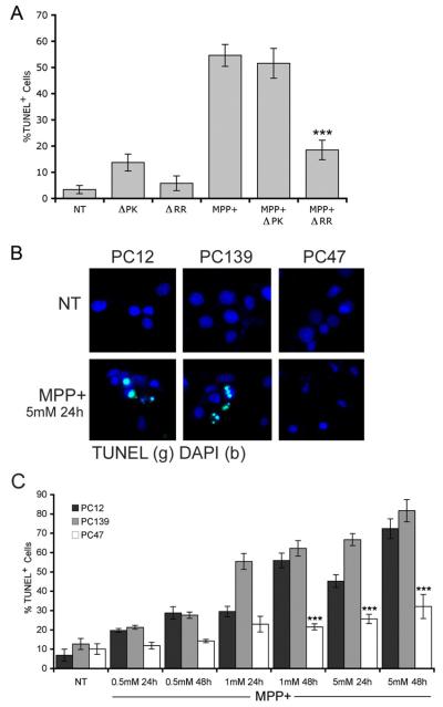
Figure 1.
PC12 cells infected with ΔRR or stably transfected with ICP10 are protected from MPP+-induced PCD. (a) PC12 cells neuronally differentiated as described in Materials and Methods were mock-infected (PBS) or infected with ΔRR or ΔPK (0.1 pfu/cell). Twenty-four hrs later they were treated with MPP+ (1 mM; 24 hrs) and examined by TUNEL as described in Materials and Methods (***, p < 0.001 relative to similarly-treated PC12 and PC139 cells). (b) Neuronally differentiatedPC12, PC139 and PC47 cells mock-treated with PBS or treated with MPP+ (5 mM; 24 hrs) were stained by TUNEL using DAPI staining to identify cell nuclei. (c) Neuronally differentiated PC12, PC139 and PC47 cells were mock-treated with PBS or treated with MPP+ (0.5, 1 or 5 mM) for 24 or 48 hrs and examined by TUNEL (***, p < 0.001 relative to similarly treated PC12 and PC139 cells). Similar results were obtained in another clonal line of ICP10PK-transfected cells (data not shown).

