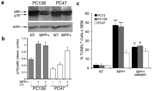
Figure 4.
MPP+-induced PCD is calpain I-dependent and inhibited by ICP10PK. (a) Extracts of neuronally differentiated PC139 and PC47 cells untreated (NT) or treated with MPP+ (5 mM, 24 hrs) were immunoblotted with antibody that recognizes full-length (80 kDa) and cleaved/activated (76 kDa) calpain I. (b) The bands from 3 independent experiments were quantitated by densitometric scanning and the p76/p80 ratios are expressed as densitometric units ± SEM. Cells treated as in (a) but in the presence of the PI3-K inhibitor LY294002 (LY, 50 μM) were similarly analyzed. PC12 cells are similar to PC139. (c) Duplicate cultures of PC12, PC139, and PC47 cells treated with MPP+ (5 mM; 24 hrs) in the presence or absence of calpeptin (50 μM) were examined for cell death by TUNEL. Results are expressed as % TUNEL+ cells ± SEM (***, p<0.001 vs. untreated; **, p<0.01 vs. MPP+).

